by Joshua Mansour, M.D.
With approximately 160,000 deaths in 2018 due to lung cancer, it is the most common cause of cancer death in the United States. The U.S Preventive Services Task Force has set out new guidelines for the use of low dose computed tomography (LDCP) that have recently been updated for individuals at high risk of having lung cancer. Lung cancer screening using this computed tomography testing has been shown to reduce death by 20-40%. However, one ongoing issue with the use of this screening exam has been the rate of false positives (a result that indicates that a person does have a disease when the actually do not). Although LDCP has helped immensely in early detection, it has been found that about one-quarter of the suspected nodules are actually not cancerous.
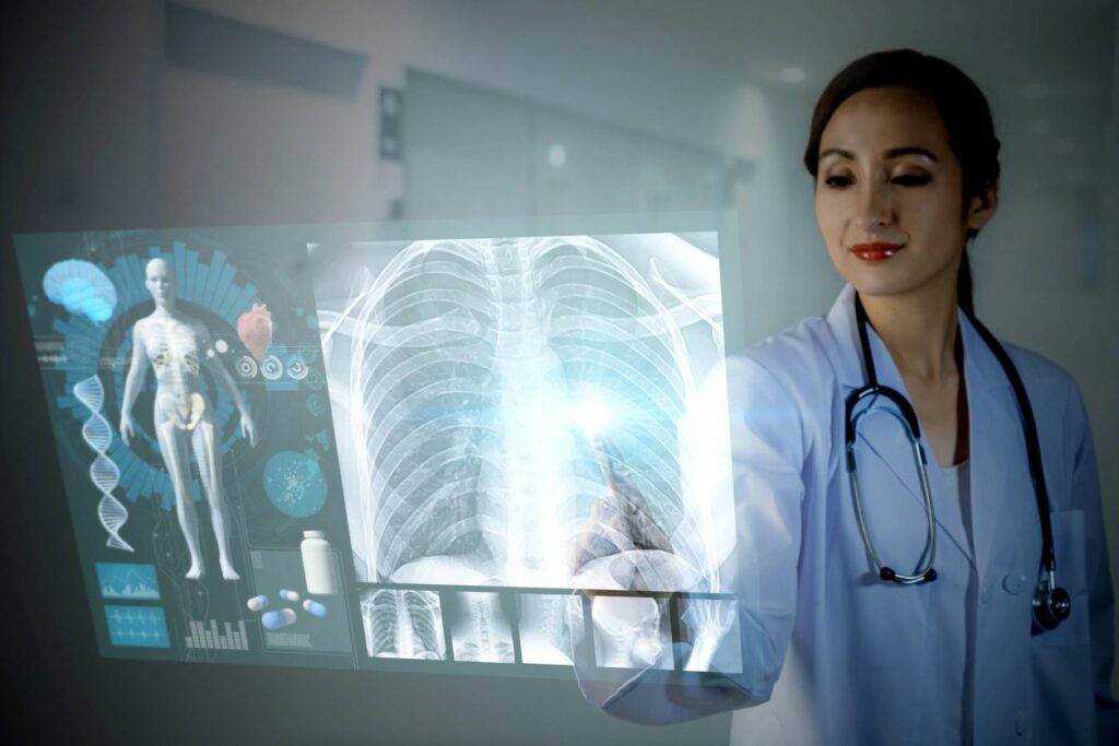
To determine if this could be improved upon, doctors at Northwestern University and Stanford, teamed up with Google to determine if a type of artificial intelligence, called Deep Learning, could help improve upon our current methods. This type of artificial intelligence essentially teaches computer systems to “learn by example”. Researchers from Google used more than 42,000 CT scans to train this the artificial intelligence to detect cancerous lung nodules on radiology imaging. The study was recently published in Nature titled “End-to-end lung cancer screening with three-dimensional deep learning on low dose chest computed tomography”. Over 6,000 National Lung Cancer Screening Trial cases were tested as well as an independent evaluation of a set of over a thousand cases in the recent study.
To understand more about this complex system, it can be broken down into three main components. The first part involves a three-dimensional model that performed analysis of whole CT scans with nodules that had been confirmed as cancerous to obtain “training data”. The second component was to train the system to detect a “region of interest” that is suspicious. Finally, the third step involves developing a cancer prediction model that would operate independently from the first two steps.
The artificial intelligence system was compared against radiologists who had evaluated low-dose chest computed tomography scans for patients, several of them who had confirmation of cancer by biopsy within a year. This deep-learning artificial intelligence system produced fewer false-negatives, as well as fewer false positives. When prior imaging was available, the model performance proves to be superior to the radiologists (six of them) with an 11% reduction in false positives and a 5% reduction in false negatives.
In most comparisons, the deep-learning system performed as well or even better than the radiologists. This study was a retrospective study that examined past cases. As pointed out by Mozziyar Etemadi, MD, Ph.D., one of the authors of the study, “the next step is to perform a prospective study to see if the tool, when used by a radiologist, can lead to earlier and more accurate diagnosis of cancer”. However, it may be some time before this can be incorporated into a hospital system. As of now the algorithm used is very sophisticated and may take some time to integrate into hospital computer systems. This is something that can be beneficial to patient care when incorporated with physicians, as both have the potential to miss something or make mistakes. The use of algorithms that may incorporate co-morbidities and risk factors in medicine is not uncommon today. However, the use of such a sophisticated one on its own will take time and prospective studies that follow patients over time and will need to be evaluated to confirm the data and include in a clinical setting. This will take a further evaluation, but there is no denying that the use of Artificial Intelligence technology continues to be an evolving asset in the medical setting
About Joshua Mansour, MD: Board-certified hematologist/oncologist working and in the field of hematopoietic stem cell transplantation and cellular immunotherapy in Stanford, California. Recently he has managed to have over 10 recent abstracts and over 10 recent manuscripts published in esteemed journals and given countless presentations at conferences and other institutions. He has helped design and implement clinical studies to evaluate current treatment plans, collaborated on grant proposals, and lead multi-institutional retrospective studies that have been published.

